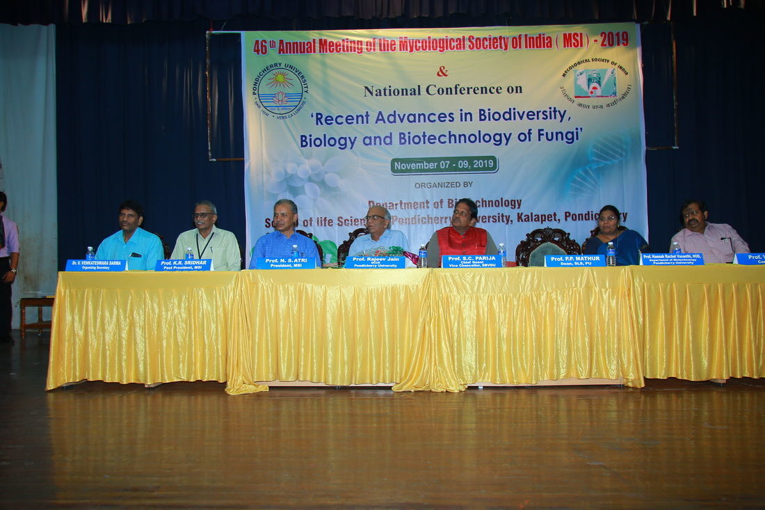
Title  |
Content |
Editorial Board |
KAVAKA 59(1): 1-6 (2023) DOI: 10.36460/Kavaka/59/1/2023/1-6
Mangrove Fungi for the Future
K. Kathiresan* and M. Kalaiselvam
Centre of Advanced Study in Marine Biology, Annamalai University, Parangipettai-608 002, India.
*Corresponding author Email:This email address is being protected from spambots. You need JavaScript enabled to view it.
(Submitted on February 22, 2023; Accepted on February 28, 2023)
ABSTRACT
Mangicolous fungi are biologically diverse and ecological important to determine the productivity of mangrove ecosystems. The mangrove fungi are largely untapped for bioprospecting potential. Further studies are required for enzymes and novel chemical entities especially glycolipids from the mangrove fungi.
Keywords: Mangroves, Mangicolous fungi, Fungal diversity, Fungal prospecting
KAVAKA 59(1): 7-15 (2023) DOI: 10.36460/Kavaka/59/1/2023/7-15
Bheemamyces uvariae sp. nov., a New Foliar Mycobiont from Andaman Archipelago, India
A. Sabeena and H. Biju*
Microbiology Division, KSCSTE - Jawaharlal Nehru Tropical Botanic Garden and Research Institute, Palode-695 562, Thiruvananthapuram, Kerala, India.
*Corresponding author Email:This email address is being protected from spambots. You need JavaScript enabled to view it.
(Submitted on November 04, 2022; Accepted on March 08, 2023)
ABSTRACT
An infrequent foliicolous fungal species, Bheemamyces uvariae sp. nov., was found infecting the leaves of Uvaria hamiltonii Hook. f. and Thomson (Annonaceae), collected from Mount Harriet National Park, South Andaman is described and illustrated in detail. Bheemamyces uvariae is characterized in having both lateral appressoria on the main hyphae and lateral, sublateral to intercalary appressoria on the hyphae originated from the main hyphae, narrower, curved, slightly elevated from the host surface and pointed at the tip. This is the first record of the genus Bheemamyces on the members of the family Annonaceae.
Keywords: Annonaceae, Bheemamyces, Black mildew, New species, South Andaman
KAVAKA 59(1): 16-24 (2023) DOI: 10.36460/Kavaka/59/1/2023/16-24
Diversity of Arbuscular Mycorrhizal Fungi in Association with Mangrove Plants in Tamil Nadu, India
C. Shankarammal and M. Kalaiselvam*
Centre of Advanced Study in Marine Biology, Faculty of Marine Sciences, Annamalai University, Parangipettai-608 002, India.
*Corresponding author Email:This email address is being protected from spambots. You need JavaScript enabled to view it.
(Submitted on February 27, 2023; Accepted on February 28, 2023)
ABSTRACT
The present study investigated the diversity of arbuscular mycorrhizal fungi (AMF) associated with four mangrove plant species viz., Avicennia marina, A. officinalis, Rhizophora apiculata and R. mucronata in two locations of Cuddalore and Mayiladuthurai districts in Tamil Nadu, India. Soil properties were analysed to determine their potential effects on the distribution of AM fungi. The present study revealed that all the mangroves had AM fungal association with varying amount of root colonization (55-86%) and soil spore density (176 to 350 spores/100g soil). Among them, R. mucronata was recorded with the maximum spore density (350 spores/100 g soil) while A. officinalis had the minimum spore density (176 spores/100 g soil). Physico chemical analyses showed the soil had slightly acidic pH (6.2-6.5), low level of phosphorus (P) (14.23-17.25 kg/acre), and high level of nitrogen (N) (51.2-54.5 kg/acre). Soil P and salinity appeared to be the important factors influencing AM fungal association in mangrove plants. The AM fungal spores of four different genera viz., Acaulospora, Gigaspora, Glomus, and Scutellospora were recorded. The AM fungi were found to be an important component on the landward fringe of mangrove habitats.
Keywords: Arbuscular mycorrhizal fungi, Acaulospora, Mangroves, Avicennia marina, A. officinalis, Gigaspora, Glomus, Scutellospora, Rhizophora apiculata, R. mucronata
KAVAKA 59(1): 25-32 (2023) DOI: 10.36460/Kavaka/59/1/2023/25-32
Talaromyces qii, a New Record of a Rare Talaromyces from the Northern Western Ghats, India
Nikhil Ashtekar1, Kunhiraman C. Rajeshkumar1,2*, Sneha Lad1, Harikrishnan K,1 and Sherin Varghese3
1National Fungal Culture Collection of India (NFCCI), Biodiversity and Palaeobiology (Fungi) Gr., Agharkar Research Institute, G.G. Agarkar Road, Pune-411 004, Maharashtra, India.
2Faculty of Science, Savitribai Phule Pune University, Ganeshkhind Rd, Ganeshkhind, Pune-411 007, Maharashtra, India.
3School of Bioscience, Mahatma Gandhi University, Kottayam-411 007, Kerala, India.
*Corresponding author Email:This email address is being protected from spambots. You need JavaScript enabled to view it.
(Submitted on December 07, 2022; Accepted on January 02, 2023)
ABSTRACT
In this study, Talaromyces qii belonging to the section Talaromyces is reported as a new record from India based on the morphology and phylogenetic analyses of four gene datasets viz. ITS, BenA, CaM, and rpb2. This is the second report of this rare Talaromyces species from across the world. Phylogenetically, the Indian strain T. qii (NFCCI 5151) formed a sister lineage to the type species T. qii (AS3 15414) due to the sequencing error in the type. The quality assessment of the four gene sequences derived from all type strains of section Talaromyces in this study evaded the proposal of a redundant novelty in this section, aligning the Indian strain NFCCI 5151 along with T. qii (AS3 15414). Morphology of type strain T. qii (AS3 15414) and T. qii (NFCCI 5151) are mostly identical, viz. elongated, biverticillate-symmetrical conidiophores, acerose phialides, and ellipsoidal or sub-globose conidia with echinulate ornamentation. However, the Indian strain has longer conidiophores and a larger conidia size than type strain T. qii and T. thailandensis. This study resolved the phylogeny of a new record of Talaromyces qii in the section Talaromyces from India through the most modern taxonomic approaches.
Keywords: Ascomycota, BenA, rpb2, Talaromyces, Trichocomaceae, India
KAVAKA 59(1): 33-47 (2023) DOI: 10.36460/Kavaka/59/1/2023/33-47
Bioprospecting Marine Fungal Enzymes-Scope and Challenges
Chandrasekaran Muthusamy* and Kalaiselvam Murugaiyan
CAS in Marine Biology Faculty of Marine Sciences, Annamalai University, Parangipettai-608 502, Tamil Nadu, India.
*Corresponding author Email:This email address is being protected from spambots. You need JavaScript enabled to view it.
(Submitted on March 04, 2023; Accepted on March 09, 2023)
ABSTRACT
Marine microorganisms are promising source of enzymes with industrial applications due to their immense genetic and biochemical diversity. Availability of novel enzymes, advancements in enzymology and enzyme technology have significantly contributed to the industrial application of enzymes and the rapid expansion of the enzyme market. In this context marine fungal enzymes assume greater attention recently owing to the great demand for novel and efficient biocatalysts for industrial applications and other services. This situation has warranted exploration of marine fungal biodiversity for new enzymes. The present review focus on bioprospecting of marine fungal enzymes produced by lesser studied fungi, identification of research gaps, challenges in pursuing research in harnessing the potentials of marine fungi, and the scope for future prospects. Role of fungal enzymes in biogeochemical processes in marine environments, bioremediation, and plastic degradation is discussed indicating marine fungi as source of industrial enzymes. Scope for exploring marine fungal diversity and potentials of extremozymes, cold adaptive enzymes and halophilic fungal enzymes, besides the need for bioprocess development are discussed. Moreover, the challenges lying ahead in pursuing research on marine fungi are also discussed to draw the attention of mycologists and biotechnologists to appropriately harness the marine fungi.
Keywords: Marine fungal enzymes, Marine fungal diversity, Potential applications, Prospects
KAVAKA 59(1): 48-55 (2023) DOI: 10.36460/Kavaka/59/1/2023/48-55
Diversity of Genus Ceriporia Donk in India
Gurpreet Kaur1, Avneet Kaur2, Ellu Ram2, Harminder Kaur2, Hardeep Kaur2, Avneet Pal Singh2* and Gurpaul Singh Dhingra2
1Department of Agriculture, Khalsa College, Amritsar-143 002, Punjab, India.
2Department of Botany, Punjabi University, Patiala-147 002, Punjab, India.
*Corresponding author Email:This email address is being protected from spambots. You need JavaScript enabled to view it.
(Submitted on May 16, 2022; Accepted on February 10, 2023)
ABSTRACT
The present paper describes eight species of the resupinate, poroid genus Ceriporia Donk based on the specimens collected from different localities in Punjab, Union Territory of Chandigarh and Himachal Pradesh (India). Of the described species, C. alachuana (Murrill) Hallenb., C. camaresiana (Bourdot and Galzin) Bondartsev and Singer, C. davidii (D.A. Reid) M. Pieri and B. Rivoire, C. microspora I. Lindblad and Ryvarden, C. reticulata (Hoffm.) Domanski, C. spissa (Schwein. ex Fr.) Rajchenb. and C. tarda (Berk.) are recorded as new to India. C. leptoderma (Berk. and Broome) Ryvarden is being reported for the first time from Punjab plains. Key to 12 species of the genus, eight described in the present paper and four reported by the earlier workers, reported so far from India is also provided.
Key words: Polypore, Mushroom, White rot, Wood rotting fungi, Pores
KAVAKA 59(1): 56-74 (2023) DOI: 10.36460/Kavaka/59/1/2023/56-74
Preparation of Nutrient Enriched Bio-Formulation(s) Using Vermi-Compost as a Carrier Material for Sustainable Agriculture
Praful Kumar*, Sandhya Sahu, and K.P. Verma
Department of Plant Pathology, Indira Gandhi Krishi Vishwavidyalaya, Raipur- 492 012, Chhattisgarh, India.
*Corresponding author Email:This email address is being protected from spambots. You need JavaScript enabled to view it.
(Submitted on November 08, 2022; Accepted on February 24, 2023)
ABSTRACT
A nutrient enriched bio-formulation(s) was prepared by inoculating the combination of bio-inoculants viz., Trichoderma sp., Pseudomonas fluorescens and Azotobacter chroococcum in vermicompost supplemented with minimal inorganic fertilizers i.e., Diammonium phosphate and Muriate of potash, which is compatible with bio-inoculants. Twenty four Trichoderma isolates were isolated from different locations of Dhamtari, Rajnandgaon, and Kabirdham District of Chhattisgarh. All the 24 Trichoderma isolates were evaluated for compatibility with P. fluorescens and A. chroococcum. Three Trichoderma isolates were found compatible with both the bacterial bio-inoculants. Trichoderma isolates TRT2, TRT-9, and TRT-12 found potentially able to produce IAA, siderophore, HCN, cellulase, chitinase, and phosphatase. Similarly, P. fluorescens and A. chroococcum individually having potential to produce IAA, siderophore, HCN, cellulase, chitinase, and phosphatase. Trichoderma isolate TRT-2 did not produce HCN and A. chroococcum did not produce cellulase enzymes. Trichoderma isolates i.e. TRT-2, TRT-9, and TRT-12, P. fluorescens and A. chroococcum individually were further evaluated for sensitivity with inorganic fertilizers i.e., DAP, MoP, SSP, urea, and complex fertilizer 28:28:00. TRT-2, TRT-9, and TRT-12 were inhibited by urea and complex fertilizer (28:28:00) at all concentrations. However, SSP could not inhibit the growth at any concentration, whereas inhibition percentages in DAP were ranged, 0.00-42.10%, 0.0-0.20%, and 0.37-19.11% for TRT-2, TRT-9, and TRT-12, respectively. Successive increases in concentration up to 5% of fertilizer could not affect the growth of isolate TRT-9 that leads to resistance. Based on the above mentioned findings, three formulations (1) Vermicompost enriched with three bio-inoculants i.e. Trichoderma isolates, P. fluorescens, and A. chroococcum, (2) DAP (5% w/w) and MoP (2% w/w) supplemented vermicompost enriched with three bio-inoculants i.e. Trichoderma isolates, P. fluorescens and A. chroococcum, and (3) DAP (10% w/w) and MoP (3% w/w) supplemented vermicompost enriched with three bio-inoculants i.e. Trichoderma isolates, P. fluorescens, and A. chroococcum, were constituted and evaluated.
Keywords: Trichoderma isolates; Pseudomonas fluorescens; Azotobacter chroococcum; Nutrient Enrichment; Bio-formulation(s).
KAVAKA 59(1): 75-82 (2023) DOI: 10.36460/Kavaka/59/1/2023/75-82
Diversity Status of Arbuscular Mycorrhizal (AM) Fungi in Association with Selected Mangrove Plants in Tamil Nadu
Anish V. Pachu and V. Mohan*
Division of Forest Protection, Institute of Forest Genetics and Tree Breeding, Coimbatore-641 002, India.
*Emeritus Professor, Centre for Advanced Studies in Botany, University of Madras, Chennai-600 025, India. *Corresponding author Email:This email address is being protected from spambots. You need JavaScript enabled to view it.
(Submitted on February 14, 2023; Accepted on February 28, 2023)
ABSTRACT
An investigation was carried out for three mangrove plant species from two coastal regions - Parangipettai and Pazhayar - of Cuddalore and Mailaduthurai districts, respectively, Tamil Nadu, India to determine their symbiotic association potential with arbuscular mycorrhizal (AM) fungi. All mangrove plants developed AM fungal colonization in their root tissues with a mean range of 85% - 95%. All the soil sediment samples had AM fungal spores with a density range from 196 - 1403 spores/100g air-dried soil sediment. Variations in AM fungal root colonization and soil spore densities were found statistically significant. Maximum percent root colonization and soil spore population of AM fungi were recorded in samples of Rhizophora mucronata in both the study locations. Frequency distribution of AM fungi was also determined, and it was found that the rhizosphere samples of three mangrove species had maximum fungal population of Rhizophora mucronata, which was followed by that of R. apiculata in both the study locations. Significance of the findings is discussed in detail.
Keywords: AM fungi, Acaulospora, Glomus, Gigaspora, Mangroves, Avicennia, Rhizophora
KAVAKA 59(1): 83-91 (2023) DOI: 10.36460/Kavaka/59/1/2023/83-91
Molecular Identification and Antagonistic Activity of Trichoderma species from Chilli Field Soil in Thiruvarur District, Tamil Nadu, India
Gomathi, S.*, Ambikapathy, V., Panneerselvam, A., and Gayathri, G.
AVVM Sri Pushpam College (Autonomous) Poondi-613 503, Thanjavur (Dt), Tamil Nadu, India.
(Bharathidasan University, Tiruchirappalli-630024, Tamil Nadu, India)
*Corresponding author Email:This email address is being protected from spambots. You need JavaScript enabled to view it.
(Submitted on February 08, 2023; Accepted on February 28, 2023)
ABSTRACT
Trichoderma asperellam is a fungal species that is frequently utilized in the biological control of plant pathogenic fungi. The creation of analytical enzymes enhances its ability to control biological infections. It has been highlighted that physical traits alone are insufficient for identifying these fungal species. As a result, the study's goal was to use molecular markers to identify Trichoderma species and assess their antagonistic effectiveness against plant pathogens. T. asperellam have antagonistic and physiologically controlled activity, which inhibited the pathogenic fungus to the greatest extent possible in a dual culture approach. T. asperellam had the highest inhibition (62.7%), followed by T. harzianum (56.0%), T. koeningii (56.0%), Aspergillus niger (52.5%), Aspergillus sulphureus (48.5%), Penicillium sp. (48.41%), and Aspergillus flavus (47.3%).
Keywords: Trichoderma sp, Biocontrol, Molecular identification, Gene sequencing
KAVAKA 59(1): 92-97 (2023) DOI: 10.36460/Kavaka/59/1/2023/92-97
Bactrospora mangrovei sp. nov., a Novel Marine Lichenized Fungus from Muthupet Mangroves of India Based on Morpho-molecular Data
B. Devadatha and V. Venkateswara Sarma*
Fungal Biotechnology Laboratory, Department of Biotechnology, School of Life Sciences, Pondicherry University, Kalapet, Pondicherry-605 014, India.
*Corresponding author Email:This email address is being protected from spambots. You need JavaScript enabled to view it.
(Submitted on March 08, 2023; Accepted on March 09, 2023)
ABSTRACT
Muthupet mangrove forests in Tamil Nadu is relatively a smaller belt when compared to other mangrove forests on the east coast of India. On 7 mangrove hosts we have recorded more than 78 fungi. The unravelling of novel marine fungi continues with the description of Bactrospora mangrovei, a new marine lichenized fungus, from the Muthupet mangroves, Tamil Nadu, East coast of India which is being reported in this paper. The species B. mangrovei is characterized by having apothecia that are numerous, frequently non-stromatic, round to irregular, coriaceous, reddish brown to black, asci bitunicate, cylindrical with short pedicels, apically rounded and ascospores uniseriate to biseriately arranged, fasciculate, filiform, 8-10 septate, partially overlapping, hyaline, rounded at both ends.
Keywords: New species, Taxonomy, Lichenized fungi, Phylogeny
Back Pages
Editors & Editorial Board
|
EDITOR-IN-CHIEF
|
|
|
Prof. Rupam Kapoor
Department of Botany, University of Delhi, Delhi-110 007, India
This email address is being protected from spambots. You need JavaScript enabled to view it.
Mobile: 91-09818497035
|
|
MANAGING EDITOR
|
|
|
Dr. Surinder Kaur Walia
Associate Professor, Department of Botany, SGTB Khalsa College, University of Delhi, Delhi - 110 007, India
This email address is being protected from spambots. You need JavaScript enabled to view it.
Mobile:+91-9811400730
|
|
ASSOCIATE MANAGING EDITORS
|
|
|
Dr. C.K. Pradeep
Senior Scientist, Jawaharlal Nehru Tropical Botanic Garden &
Research Institute
Thiruvananthapuram- 695 562, India
This email address is being protected from spambots. You need JavaScript enabled to view it.
This email address is being protected from spambots. You need JavaScript enabled to view it. +91- 9446849654
|
|
Dr. V. Venkateswara Sarma
Assistant Professor, Dept. of Biotechnology, School of life Sciences, Pondicherry University
Pondicherry – 605014, India
This email address is being protected from spambots. You need JavaScript enabled to view it.
+91- 9444467787
|
|
SECTION EDITORS
|
|
Fungal Systematics, Evaluation, Phylogeny and Evolution
|
|
|
Prof. Yashpal Sharma
Department of Botany, University of Jammu, Jammu -180 006, India
+91-9419157412
This email address is being protected from spambots. You need JavaScript enabled to view it.
|
|
Prof. Krishnendu Acharya
Department of Botany,
University of Calcutta, Kolkata-700 019, West Bengal, India
This email address is being protected from spambots. You need JavaScript enabled to view it.
+91 8013167310
|
|
Mycorrhiza, Lichens and related Fungi
|
|
|
Prof. B.F. Rodrigues
Head, Department of Botany, Goa University
Goa- 403 206, India
This email address is being protected from spambots. You need JavaScript enabled to view it.
+91 9422446359
|
|
Dr. Sanjeeva Nayak
Principal Scientist,
Lichenology Lab,
CSIR-National Botanical Research Institute, Lucknow-226 001, India
This email address is being protected from spambots. You need JavaScript enabled to view it.
+91 8756104655
|
|
Fungal Biotechnology
|
|
|
Prof. J. Savitha
Head, Department of Microbiology & Biotechnology, Bangalore University, Jnanabharathi Campus Bangalore-560 056, India
This email address is being protected from spambots. You need JavaScript enabled to view it.
+91-9886185170
|
|
Prof. Naveen Kango
Head, Department of Microbiology,
Dr. Harisingh Gour Vishwavidyalaya, Sagar - 470003,
Madhya Pradesh, India
This email address is being protected from spambots. You need JavaScript enabled to view it.
+91-9425635736
|
|
Marine Biology and Fungal Physiology
|
|
|
Prof. M. Kalaiselvam
Director, CAS in Marine Biology, Faculty of Marine Sciences,Annamalai University. Parangipettai – 608 502, India
This email address is being protected from spambots. You need JavaScript enabled to view it.
Mobile: +91-9894445103
|
|
Prof. N. Thajuddin
Head, Department of Microbiology,
Bharathidasan University,
Tiruchirappalli – 620 024, India
This email address is being protected from spambots. You need JavaScript enabled to view it.
+91 9842379719
|
|
Plant Pathology and Disease Management
|
|
|
Prof. N. Mathivanan
Director, Centre for Advanced
Studies in Botany, University of Madras, Guindy Campus, Chennai - 600 025, India
This email address is being protected from spambots. You need JavaScript enabled to view it.
+91-9840253789
|
|
Dr. Pankaj Baiswar
Principal Scientist, Plant Pathology, ICAR Research Complex, For NEH Region, Umiam – 793103, India
This email address is being protected from spambots. You need JavaScript enabled to view it.
+91 9436107733
|
|
Editorial Board
|
|
|
Prof. Resurreccion B. Sadaba
Division of Biological Sciences, College of Arts and Sciences,
University of the Philippines Visayas, Philippines.
This email address is being protected from spambots. You need JavaScript enabled to view it.
|
|
Prof. D.J. Bagyaraj
NASI Senior Scientist & Chairman, Centre for Natural Biological Resources and Community Development, 41, RBI Colony, Anand Nagar, Bangaluru – 560 024, India.
This email address is being protected from spambots. You need JavaScript enabled to view it.
+91-9448465368
|
|
|
Prof. T.N. Lakhanpal
Department of Biosciences H.P. University, Shimla-171 005, India.
This email address is being protected from spambots. You need JavaScript enabled to view it.
+91-9816264141
|
|
Prof. C. Manoharachary
NASI Senior Scientist
Department of Botany, Osmania University,
Hydrabad-500 007, Andhra Pradesh
This email address is being protected from spambots. You need JavaScript enabled to view it.
+91-93911 64243
|
|
|
Prof. D.J. Bhat
128/1-J, Azad Housing Society, Curca, P.O. Goa Velha, Goa -403108, India
This email address is being protected from spambots. You need JavaScript enabled to view it.
+91-9890946529
|
|
Dr. Chandralata Raghukumar
Emeritus Scientist
National Institute of Oceanography,
Dona Paula, Goa -403 004
This email address is being protected from spambots. You need JavaScript enabled to view it.
+91-9890404044
|
|
|
Prof.T. Satyanarayana
Department of Microbiology, University of Delhi,
South Campus, Benito Juarez Road, New Delhi -110 021, India.
This email address is being protected from spambots. You need JavaScript enabled to view it.
Tel:91-011-24112008
|
|
Prof. N.S. Atri,
Department of Botany, Punjabi University, Patiala-147 002, India.
This email address is being protected from spambots. You need JavaScript enabled to view it., This email address is being protected from spambots. You need JavaScript enabled to view it.
+91-9417810754
|
|
|
Prof. Geeta Sumbali
Department of Botany,
University of Jammu,
Jammu -180 006
gThis email address is being protected from spambots. You need JavaScript enabled to view it.
+91 9419192102
|
|
Prof. M.V. Rajam
Department Department of Genetics, University of Delhi, South Campus, DhaulaKuan, New Delhi 11002, India
This email address is being protected from spambots. You need JavaScript enabled to view it.
+91-9818108515
|
|
|
Prof. K.R. Sridhar
Department of Biosciences, Mangalore University, Mangalagangotri, Mangalore 574 199, India
This email address is being protected from spambots. You need JavaScript enabled to view it.
+91-9741993216
|
|
Prof. K.R. Aneja
Chairman, Department of Microbiology
Kurukshetra University
Kurukshetra -136 119, Haryana
anejakr@yahoo.ca
Mobile: +91-9466241532
|
|
|
Prof. N.K. Dubey
Botany Department,
Banaras Hindu University Varanasi-221 005, Uttar Pradesh
This email address is being protected from spambots. You need JavaScript enabled to view it.
+91-9415295765
|
|
Prof.Absar Ahmad
Director, Interdisciplinary Nanotechnology Centre,
Aligarh Muslim University, Aligarh-202001, India
This email address is being protected from spambots. You need JavaScript enabled to view it.
Mobile : +91-9823064820
|
|
|
Prof. Munruchi Kaur
Department of Botany, Punjabi University, Patiala – 147002, India
This email address is being protected from spambots. You need JavaScript enabled to view it.
|
|
Dr. Sanjay K. Singh
Scientist, National Fungal Culture Collection of India,
MACS Agharkar Research Institute, GG Agarkar Road,
Pune – 411004, India
This email address is being protected from spambots. You need JavaScript enabled to view it.
+91-9423239056
|
|
|
Dr. K. B. Vrinda
Jawaharlal Nehru Tropical Botanic Garden & Research Institute,
Palode, Thiruvananthapuram- 695 562, India
This email address is being protected from spambots. You need JavaScript enabled to view it.
+91 9447208372
|
|
Prof. R.M. Mulani,
Professor of Botany & Director, School of Life Sciences,
Swami Ramanand Teerth Marathwada University, Nanded -431606, Maharashtra
This email address is being protected from spambots. You need JavaScript enabled to view it.
+91 9657224391, +91
|
|
|
Prof. Ashish Vyas,
Head, Department of Microbiology, School of Biotechnology & Biosciences,
Lovely Professional University, Phagwara – 144411, India
This email address is being protected from spambots. You need JavaScript enabled to view it.
+91 7696660630
|
|
Dr. Kanad Das
Scientist-E, Botanical Survey of India, Crpytogamic Unit,
P.O. Indian Botanic Garden, Howrah- 711 103, India
This email address is being protected from spambots. You need JavaScript enabled to view it.
Mobile : +91 9932147368
|
|
|
Dr. Sushil Kumar Shahi
Associate Professor,
Botany Department,
Guru Ghasidas Vishwavidyalaya,
Bilaspur - 495009, India
This email address is being protected from spambots. You need JavaScript enabled to view it.
Mobile : 9479230418
|
|
Dr. P.Y. Prakash,
Associate Professor and In-charge Medical Mycology Division, Dept. of Microbiology, 1st Floor, Centre for Basic Sciences
Kasturba Medical College, Manipal Academy of Higher Education, Madhavnagar, Manipal-576 104, India
This email address is being protected from spambots. You need JavaScript enabled to view it.
91 988 661 6153
|
|
|
Dr. K.P. Kannan
Associate Professor, Dept. of Biotechnology,
Bannari Amman Institute of Technology, Sathyamangalam - 638 401, India
This email address is being protected from spambots. You need JavaScript enabled to view it.
+91- 9003457744
|
|
Dr. Yogesh Joshi
Associate Professor, Department of Botany, Central University of Rajasthan SSJ Campus, Almora- 263601, India
This email address is being protected from spambots. You need JavaScript enabled to view it.
Mobile : +91 9415760604
|
| Title |
Content |
Editorial Board |
KAVAKA 58 (4): 1-9 (2022) DOI: 10.36460/Kavaka/58/4/2022/1-9
Understanding onychomycosis - a neglected form of cutaneous mycosis
Geeta Sumbali* and Anjali Sharma
Department of Botany, University of Jammu, BR Ambedkar Road, Jammu-180006 (India) *Corresponding author Email: This email address is being protected from spambots. You need JavaScript enabled to view it.
(Submitted on May 27, 2022; Accepted on October 03, 2022)
ABSTRACT
Onychomycosis refers to the chronic fungal infection of toenails and fingernails that may involve any component of the nail unit. It is the most common nail disorder experienced worldwide and is usually caused by different species of dermatophytes, saprophytic moulds, yeast and yeast-like fungi. Approximately 50-70 per cent of the nail disorders are caused by fungal pathogens, which affect the quality of life by causing pain, discomfort and physical impairment. Moreover, onychomycosis cause psychological and social limitations, which can undermine work and social life of a person. Onychomycosis is mostly caused by keratinophilic fungi, which have the unique ability of degrading keratin that forms the nail apparatus. Structurally, the nail apparatus has certain protective layers, such as, the cuticle and the distal solehorn but it is exposed to the harsh environments and is thus prone to damage and invasion by various keratinophiles through the distal and proximal nail folds. Management of onychomycosis is quite challenging since the infection is embedded within the nail and is difficult to reach. Synthetic antifungal agents are commonly used to control them but they are non-biodegradable, have various side effects due to residual toxicity, long duration of treatment, recurrence of infection and development of resistance to fungal pathogens. All these reasons have led to the exploration of some naturally occurring fungitoxicants like essential oils of higher plants for the management of onychomycotic pathogens
Keywords: Onychomycosis, Nail dystrophies, Dermatophytes, Non-dermatophytic filamentous fungi, Yeast-like fungi
KAVAKA 58 (4): 10-17 (2022) DOI: 10.36460/Kavaka/58/4/2022/10-17
Eco-fabrication of anti-proliferative fluorescent silver nanoparticles using neem endophytic fungus
Ejaz Ahmad Siddiqui1, Shadab Khan1, Sahrish Arfin1, Shruti Satpute1, Pooja Salunkhe1, Iqbal Siddiqui1, Sk Najrul Islam2, Renuka Bhor3, Jyoti Otageri3, Kalpana Pai3, Narendra Kadoo3, Vidya Gupta3 and Absar Ahmad1,2*
1Division of Biochemical Sciences, CSIR-National Chemical Laboratory, Pune-411008, India 2Interdisciplinary Nanotechnolcogy Centre (INC), Z.H. College of Engineering and Technology, Aligarh Muslim University, AMU, Aligarh, UP-202002, India.
3Department of Zoology, Savitribai Phule Pune University, Pune -411007, India. *Corresponding author Email: This email address is being protected from spambots. You need JavaScript enabled to view it.
(Submitted on September 22, 2022; Accepted on December 14, 2022)
ABSTRACT
Ever since scientists uncovered that plant-associated endophytic fungi have the ability to autonomously generate the same plant-based drugs/bioactive molecules; a multitude of endophytes have been utilized for the same. Extending the above aspect, our group directed it towards employing endophytes instead of plants in the 'eco-fabrication of nanoparticles'. The present manuscript demonstrates one such simple, reliable and ecological approach to the synthesis of biomedically and technologically prized silver nanoparticles (AgNPs) using an endophytic fungus isolated from neem (Azadirachta indica A. Juss.) leaves. Wet mycelial mass of the fungus when added to aqueous precursor silver nitrate (AgNO3) led to the synthesis of profuse amounts of extracellular dispersed, fluorescent, quasi-spherical AgNPs of 20-60 nm having 20 nm average size. The nanoparticles were utterly characterized using recognized standard techniques. Cytotoxic activity of these nanoparticles was examined against human lung cancer cells (A549) and normal human peripheral blood mononuclear cells (PBMC), and it was observed that our AgNPs are anti-proliferative against cancer cells but safe toward normal cells. Furthermore, assessment of toxicity toward human RBCs (erythrocytes) revealed a mere 7% hemolysis in comparison to Triton X-100, consequently confirming the safe nature of our nanoparticles on human cells. Also, we scrutinized the anti-microbial potency of our biofabricated AgNPs and found them to be anti-microbial against different fungal (Aspergillus niger) and bacterial [Gram positive (Bacillus subtilis & Staphylococcus aureus), Gram negative (Pseudomonas aeruginosa)] strains. These multi-faceted nanoparticles will find broad spectrum applications in fields like drug/targeted delivery, therapeutics, theranostics, anti- microbial coatings, and so on
Keywords: Anti-microbial, Anti-proliferative, Eco-fabrication, Endophyte, Neem, Silver nanoparticles
KAVAKA 58 (4): 18-23 (2022) DOI: 10.36460/Kavaka/58/4/2022/18-23
Morphological and phylogenetic characterisation of two species of family Russulaceae from Jammu and Kashmir, India
Komal Verma1, Faisal Mushtaq2, Anil Kumar1 and Yash Pal Sharma1*
1Department of Botany, University of Jammu, Jammu, Jammu and Kashmir, India. 2Department of Botany, Lovely Professional University, Punjab, 144001, India. *Corresponding author Email: This email address is being protected from spambots. You need JavaScript enabled to view it.
(Submitted on August 17, 2022; Accepted on November 03, 2022)
ABSTRACT
In the present paper two species of family Russulaceae i.e., Lactarius abieticola and Russula lakhanpalii are reported for the first time from Jammu and Kashmir, India. Russula lakhanpalii belongs to subgenus Heterophyllidia of genus Russula and Lactarius abieticola belongs to subg. Lactarius of genus Lactarius. A detailed macro- and micromorphological descriptions coupled with the illustrations and nrITS-based molecular analyses are presented here.
Keywords: Macrofungi, nrITS, Phylogeny, Regional records
KAVAKA 58 (4): 24-27 (2022) DOI: 10.36460/Kavaka/58/4/2022/24-27
Effect of native rice specific isolates of Trichoderma and ecological fitness against aggregated sheath spot of rice caused by Ceratorhiza oryzae-sativae
Konjengbam Sarda Devi1*, Kota Chakrapani1, Wangkhem Tampakleima Chanu1, Bijeeta Thangjam1, Bireswar Sinha1, Laikangbam Nongdrenkhomba1, Ph. Sobita Devi1 and Akoijam Ratankumar Singh2
1Department of Plant Pathology, College of Agriculture, Central Agricultural University (Imphal), Iroisemba- 795 004, Imphal West, Manipur, India
2ICAR Research Complex for North Eastern Hill Region, Manipur Centre, Lamphelpat, Imphal West-795 004, Manipur, India
*Corresponding author Email: This email address is being protected from spambots. You need JavaScript enabled to view it.
(Submitted on October 05, 2022; Accepted on December 06, 2022)
ABSTRACT
Trichoderma is a free-living fungus that interacts heavily with its surroundings found in the root, soil, and foliar regions of plants as well. It is an important biocontrol agent due to its abilities, such as mycoparasitism, production of antibiotic, hydrolytic enzymes, competition for nutrients, as well as induced plant resistance; production of numerous secondary metabolites inhibiting the growth of several plant pathogens. Antagonistic potential of fourteen (n=14) native rice specific Trichoderma isolates was evaluated against aggregated sheath spot of rice caused by Ceratorhiza oryzae-sativae. It revealed that all native Trichoderma isolates significantly inhibited the mycelial growth of the pathogen of C. oryzae-sativae with ranges from 71.50% to 97.50% with the highest per cent inhibition by T. harzianum (MH257323), and the least percent inhibition by T. koningiopsis (MN080228). Bell's scale studied showed that class III category by T. koningiopsis (MN080228) and class II showed by T. harzianum (MH257323) against C. oryzae-sativae. Among isolates of native rice specific T. harzianum, MH257323 is found to be the most effective in reducing the rapid growth of pathogen and having high potential ecological fitness.
Keywords: Aggregated sheath spot, Biocontrol agent, Ecological fitness, Mycoparasitism, Rice, Trichoderma
KAVAKA 58 (4): 28-35 (2022) DOI: 10.36460/Kavaka/58/4/2022/28-35
Effect of AM fungus on phosphorus nutrition of maize and pigeon pea in alfisols as influenced by different phosphorus amendments of North Carolina Rock Phosphate (NCRP)
R. Mythra*, G.S. Srikanth., U. Jagadeesh, K.B. Bhagyashree and A. Manjunath
Department of Agricultural Microbiology, GKVK, University of Agricultural Sciences, Bengaluru, Karnataka, India
*Corresponding author Email: This email address is being protected from spambots. You need JavaScript enabled to view it.
(Submitted on September 19, 2022; Accepted on November 30, 2022)
ABSTRACT
A greenhouse investigation was conducted to determine the effect of AM fungus on phosphorus nutrition of maize and pigeon pea in alfisols as influenced by different phosphorus amendments of North Carolina Rock Phosphate (NCRP). This experiment consisted of 24 treatments resulting from factorial combination of two plant species, two levels of AM fungal (Glomus aggregatum) inoculation and six levels of phosphorus amendments. The extent of mycorrhizal fungal colonization in roots of pigeon pea was higher than that of maize. Inoculation of soil with AM mycorrhizal fungus caused significant increase in total phosphorus uptake of pigeon pea as well as maize. The extent of increase in total phosphorus uptake due to mycorrhizal inoculation in pigeon pea was higher than maize. The phosphorus uptake efficiency of maize was lower than pigeon pea. Mycorrhizal colonization significantly reduced phosphorus utilization efficiency of both plant species. The results of this study suggest that application of NCRP bio-acidulated with Bacillus sp. and Penicillium sp. improves phosphorus nutrition of maize and pigeon pea and inoculation of soil with AM fungus increase that effect.
Keywords: AM fungi, NCRP, Glomus aggregatum, Bacillus sp., Penicillium sp., Phosphorus nutrition.
KAVAKA 58 (4): 36-39 (2022) DOI: 10.36460/Kavaka/58/4/2022/36-39
Efficacy of fungicides and endophytic bacteria against Fusarium wilt of chickpea caused by Fusarium oxysporum f. sp. ciceris under in vitro conditions
Kailash Patel1*, R.K. Tombisana Devi1, A. Ratankumar Singh2, Kennedy Ningthoujam1, Veronica Kadam1 and Sushanti Thokchom1
1School of Crop Protection, College of Post Graduate Studies in Agricultural Sciences, CAU(I), Umiam-793103, Meghalaya, India
2Division of Crop Science, ICAR Complex for North Eastern Hill Region, Umiam-793 103, Meghalaya, India
*Corresponding author Email: This email address is being protected from spambots. You need JavaScript enabled to view it.
(Submitted on October 08, 2022; Accepted on December 09, 2022)
ABSTRACT
Chickpea (Cicer arietinum L.) is an important leguminous crop originated from Southwestern Asia region and Fusarium wilt disease caused by Fusarium oxysporum f. sp. ciceris (Foc) is limiting factor in chickpea cultivation. Efficacy of fungicides and endophytic beneficial bacteria were tested in vitro against chickpea wilt pathogen. Four different fungicides namely, carbendazim 50% WP (bavistin), hexaconazole 5% EC (contaf plus), propiconazole 25% EC (Tilt) and thiophanate methyl 70% WP (Roko) at 0.10, 0.15 and 0.20% were evaluated in vitro against Fusarium wilt of chickpea. Carbendazim and propiconazole proved the most effective exhibiting mean mycelial growth inhibition of 100% at all concentrations followed by hexaconazole and thiophanate methyl inhibit mycelial growth of 78.35 and 77.25% at 0.20% respectively. Four endophytic bacterial strains of Bacillus designated as ECP1 (Bacillus cereus), ECP5 (Bacillus subtilis), ECP8 (Bacillus amyloliquefaciens) and ECP10 (Bacillus cereus) were also evaluated against the Foc. The endophytic Bacillus strains revealed that ECP1 is the most efficacious resulted in 69.62 % mean inhibition of mycelial growth followed by ECP5 with 67.03%. ECP8 (Bacillus amyloliquefaciens) and ECP10 (Bacillus cereus) showed 65.18% growth reduction over control respectively.
Keywords: Bacillus, Chickpea, Endophytes, Fungicides, Fusarium oxysporum f. sp. ciceris
KAVAKA 58 (4): 40-46 (2022) DOI: 10.36460/Kavaka/58/4/2022/40-46
Fungal pathogens as potential mycoherbicides to control water hyacinth (Eichhornia crassipes)
K.R. Aneja
Department of Microbiology, Kurukshetra University, Kurukshetra, Haryana 136 119, India Corresponding author Email: This email address is being protected from spambots. You need JavaScript enabled to view it.
(Submitted on July 12, 2022; Accepted on October 18, 2022)
ABSTRACT
Water hyacinth (Eichhornia crassipes), a free-floating aquatic weed and native of Amazon River, is one of the fastest growing plants whose seeds can remains viable for more than 28 years in the mud. Controlling methods for water hyacinth include physical, chemical and biological, but biological using fungal pathogens and/or insects or both as a consortium is effective and eco-friendly. The world of fungi provides a fascinating and almost endless source of biological diversity, which is a rich source for exploitation. Studies conducted on fungal diversity of water hyacinth yielded 21 fungal pathogens, of these two: Fusarium chlamydosporum and Bipolaris sorokiniana recorded for the first time on this weed globally and 7 fungal pathogens [Alternaria alternata isolate-1 and isolate-2 (AL-14), Cercospora rodmanii, Phoma sorghina, Epicoccum nigrum, Acremonium sp., Alternaria sp. and Stemphylium sp.] identified as new records for the country. Biocontrol studies conducted on seven fungal pathogens showed maximum biocontrol efficacy in Cercospora rodmanii (95.3%), followed by Alternaria eichhorniae (80%) and A. alternata (45-72%), revealing all the desired characteristics, such as: can be easily cultured and maintained on natural host, host -specific, easily disseminated and can be mass produced in a short span of time. Moreover, the phylloplane microflora is not antagonistic to these pathogens, thus the biocontrol efficacy would not be affected by the surface microflora. They have the potential to be developed as a mycoherbicide/s either alone or as a consortium to manage water hyacinth worldwide either used alone or in combination with insects and or herbicide.
Keywords: Water hyacinth, Bioherbicides, Alternaria alternata, A. eichhorniae, Fusarium chlamydosporum, Epicoccum nigrum, Cercospora rodmanii
KAVAKA 58 (4): 47-50 (2022) DOI: 10.36460/Kavaka/58/4/2022/47-50
New records of lichens (Lichenized Ascomycota) from India with novel habitat preferences
Rajesh Bajpai1,2*, Ramya Ranjan Paul2, Chandra Prakash Singh3, Anzar A. Khuroo4 and Dalip Kumar Upreti2
1Environment, Agriculture and Education Society, Bareilly-234001, India
2Plant Diversity Systematics and Herbarium Division, CSIR-National Botanical Research Institute, Lucknow-226001, Uttar Pradesh, India.
3AED//BPSG/EPSA, Space Applications Centre-ISRO, Ahmedabad, India
4Centre for Biodiversity & Taxonomy, Department of Botany, University of Kashmir, Srinagar- 190006, India
*Corresponding author Email: This email address is being protected from spambots. You need JavaScript enabled to view it.
(Submitted on September 12, 2022; Accepted on November 05, 2022)
ABSTRACT
The present paper reports seven species of lichens for the first time from India. These include Cryptothecia aleurinoides Aptroot & Wolseley, Cryptothecia genuflexa (Müll. Arg.) R. Sant., Cryptothecia scriblitella (Nyl.) Makhija & Patw., Dioryogma upretii Sipman, Distothelia rubrostoma (Aptroot) Aptroot & Lücking, Umbilicaria leiocarpa DC., and Verrucaria adelminienii Zschacke. The species are appended here with brief description, distribution, ecology and specimen examined details. Most of the species prefer to grow on tree bark.
Keywords: Ascomycota, Biodiversity, Lichenized fungi, Taxonomy, India
KAVAKA 58 (4): 51-55 (2022) DOI: 10.36460/Kavaka/58/4/2022/51-55
Eco-friendly management of sheath blight in rice under organic conditions
R. Gopi1*, R.K. Avasthe2, C. Kapoor3, H. Kalita4 and S.K. Das5
1ICAR-Sugarcane Breeding Institute, Research Centre, Kannur, Kerala-670002
2National Rainfed Area Authority, Ministry of Agriculture &Farmers Welfare, Govt. of India, PUSA, New Delhi-110012
3Indian Agricultural Research Institute, PUSA, New Delhi- New Delhi-110012
4ICAR Research Complex for NEH Region, Arunachal Pradesh Centre, Basar-791101
5ICAR Research Complex for NEH Region, Sikkim Centre, Tadong, Gangtok- 737 102 *Corresponding author Email: This email address is being protected from spambots. You need JavaScript enabled to view it.
(Submitted on September 27, 2022; Accepted on December 06, 2022)
ABSTRACT
Sheath blight is the most important diseases in rice. In the study, various eco-friendly treatments were attempted to manage sheath blight in rice both in vitro and in vivo conditions. Among the various Trichoderma isolates tested, the isolate T2 collected from Pangthang (27.73° N, 88.63° E) was very effective in inhibiting the growth of sheath blight pathogen with 78.88 per cent inhibition over control and among the plant extracts fern leaves (5%)+garlic bulb (5%)+neem leaves (5%) extract combination @ 15% showed maximum percent inhibition (74.44%) over control. All the fungicides tested under field conditions performed well and better than biocontrol agents and botanicals. Among treatments, copper oxychloride @ 0.25%, was effective in the variety PD 10 with no incidence of disease. Similar result was also observed in the variety TN 1 in which copper oxychloride @ 0.25% registered lowest PDI of 7.41 per cent. Among the biocontrol agents Pseudomonas fluorescens recorded less PDI i.e 27.77 per cent and 26.65 per cent, respectively in PD 10 and TN 1.
Keywords: Disease, Eco-friendly, Rice, Sikkim, Rhizoctonia solani
KAVAKA 58 (4): 56-59 (2022) DOI: 10.36460/Kavaka/58/4/2022/56-59
New record of Choanephora cucurbitarum (Berk & Ravenel) Thaxt. infecting Colocasia esculenta ‘Fontanesii’ and other hosts from Chhattisgarh
Harvinder Kumar Singh1*, Pradeep Kumar Badhai1, Anurag Kerketta2, A. S. Kotasthane1 and C.S. Shukla1
1Department of Plant Pathology, College of Agriculture, IGKV, Raipur, Chhattisgarh, India 492012
2College of Horticulture and Research Station, Jagdalpur, IGKV, Chhattisgarh, India 494001 *Corresponding author Email: This email address is being protected from spambots. You need JavaScript enabled to view it.
(Submitted October 07, 2022; Accepted on November 29, 2022)
ABSTRACT
Choanephora cucurbitarum is a facultative saprophyte fungus belongs to phylum Zygomycota and causes fruit rot, flower rot and leaf blight disease in several host plants. During the rainy season of 2017-18, infections were observed from different locations in Chhattisgarh in Colocasia esculenta, Brassica olearcea var. botrytis, Lablab purpureus, Abelmoschus esculentus, Solanum melongena, Lagenaria siceraria, Capsicum annum and Solanum lycopersicum. Water-soaked necrotic spots on leaves later necrotic spots coalescing and blighting on leaves. On severe infection leaf lamina reduced to rotten pulpy mass. Silvery spine or whiskers like sporangiophore bearing dark spores (superficial sporangia) on spadix coupled with black rot and dieback of the flower of colocasia, black rot in fruits of brinjal, necrosis in cauliflower, necrosis on fruits of hyacinth Bean, flowers of okra, flowers of bottlegourd, fruits of chili and fruits of tomato. Sporangium was terminal and usually pendent on the recurved end of an erect sporangiophore with a definite columella which was globose in shape. Aseptate branches further swell to form young ampulla bearing spores. The disease was symptomatically and microscopically confirmed with earlier published standard monographs and literature.
Keywords: Choanephora, Colocasia, Flower rot, Wet rot.
KAVAKA 58 (4): 60-75 (2022) DOI: 10.36460/Kavaka/58/4/2022/60-75
Antifungal proteins: An ecofriendly approach for sustainable alternative of biocontrol against the disease-causing agents in plants
Praveen Gehlot1*, Dilip Singh Solanki1, Alkesh Tak1, Kamna Sharma1 and Sunil Choudhary2 1Department of Botany, Jai Narain Vyas University, Jodhpur, India-342 001.
2SBRM Government PG College, Nagaur, India- 341001 *Corresponding author Email: This email address is being protected from spambots. You need JavaScript enabled to view it.
(Submitted on September 29, 2022; Accepted on December 03, 2022)
ABSTRACT
The chemical fungicides applied to counter the diseases associated with annual and perennial crops are creating a major concern by affecting the environment adversely. Furthermore, improper and inadequate application of these fungicides leads to a process of co-evolution that develops resistance in fungal pathogens against these compounds. This current scenario has aggravated the search for alternative disease management strategies and/or safer antifungal agents that could substitute the current fungicides with bio-fungicides. Fungicides of biological origin are the botanicals proteins remain present in different plant parts and seems to be involved in either constitutive or induced resistance to pathogenic fungal attack and thus play a vital role in plant defense system against pathogenic fungi through controlling their spread. A great number of antifungal peptides and proteins have already been reported, with more are being discovered almost daily. Till now, 17 families of antifungal proteins have been identified that have a high potential for therapeutic applications in agriculture for biocontrol of pathogenic microbes that protect plants against diseases.
Keywords: Antifungal proteins, Bio-pesticides, Biological control, Fungicide
KAVAKA 58 (4): 76-85 (2022) DOI: 10.36460/Kavaka/58/4/2022/76-85
Rationally designed peptide for rapid detection of ochratoxin in paper-based dot-blot assay and mitigation of toxicity
Shraddha Rahi and Vandana Ghormade*
Nanobioscience Group, Agharkar Research Institute, Pune-411004
*Corresponding author Email: This email address is being protected from spambots. You need JavaScript enabled to view it.
(Submitted on September 07, 2022; Accepted on December 04, 2022)
ABSTRACT
Ochratoxin A (OTA) is a secondary metabolite produced by the Aspergillus spp. that contaminates a variety of food and feed. OTA is a nephrotoxic, hepatotoxic and carcinogenic mycotoxin. Here we developed a dot-blot assay for OTA detection using a specific 9-mer N- KSGSFNHPK-C peptide designed by rational strategy and computational modelling. The peptide binding to OTA was confirmed by fluorescence quenching (1.1 – 1.5-fold). The 9-mer peptide was conjugated to gold nanoparticles as labels for use as a detection agent in the dot- blot assay. The limit of detection for the developed dot-blot assay was 0.19 μg/kg. Further, OTA recovery from spiked wheat sample by dot-blot assay (90-96 %) was comparable to HPLC method (97 – 99 %). Furthermore, evaluation of developed assay with 146 samples of food and feed along with certified reference material demonstrated good sensitivity (74 %) and specificity (99 %), respectively. The assay displayed 94 % assay accuracy with correlation of 0.75 with HPLC. Moreover, the addition of peptide reduced OTA toxicity (21 – 60 %) to HepG2 cells. Therefore, an easy-to-use, rapid, and portable dot- blot assay can contribute in the sensitive detection of OTA and has potential for reducing the toxicity of OTA and ensures safe and healthy food.
Keywords: Ochratoxin, Gold nanoparticles, Rational design peptide, Dot-blot assay
Back Pages
| Title |
Content |
Editorial Board |
KAVAKA 58 (3): 1-10 (2022)
Immunodetection of Rhizophagus fasciculatus and Gigaspora gigantea in soil and root tissues in Citrus reticulata, their exploitation as bioinoculants and cellular localization of defense enzymes following induced immunity developed against Fusarium solani
B.N. Chakraborty1* and Sanjita Allay2
1Department of Biological Sciences, Aliah University, New Town, Kolkata 700 016
2Department of Botany, Sambhu Nath College, Labpur 731303
*Corresponding author Email: This email address is being protected from spambots. You need JavaScript enabled to view it.
(Submitted on July 15, 2022; Accepted on September 05, 2022)
ABSTRACT
Two dominant arbuscular mycorrhizal (AM) fungi Rhizophagus fasciculatus and Gigaspora gigantea, their colonization with root tissues in Citrus reticulata along with their scanning electron microscopic views have been presented. Immunological formats for the detection of these two AM fungi were developed. IgG raised against R. fasciculatus and Gi. gigantea were used for immunodetection of AM fungal spores in soil labeled with FITC conjugates following an indirect immunofluorescence test. AM fungal spores showed a bright apple green fluorescence which was distributed throughout the spore wall. Subtending hyphae also gave apple green fluorescence. Spores with their hyphae were more prominent in the rhizosphere. Ultrathin sections of AM fungal colonized root stained with toluidine blue confirmed the presence of fine arbuscule branches within the root cells. Immunogold localization of AM fungi in mandarin roots was demonstrated. The gold particles were mostly concentrated near the cell wall. R. fasciculatus and Gi. gigantea were tested singly and in combination for their effect in inhibiting root rot of mandarin seedlings caused by F. solani in field conditions. Joint inoculation with AMF could effectively reduce disease incidence, correlated with increased accumulation of defense enzymes such as chitinase, b-1,3-glucanase, peroxidase, and phenylalanine ammonia-lyase. Cellular localization of chitinase in mandarin root and leaf tissues have been demonstrated following indirect immunofluorescence test using PAb raised against chitinase and labeled with FITC. Immunogold localization of chitinase following immunity induced by AM fungi in mandarin plants against F. solani confirmed the immunofluorescence results. It precisely showed the sites of chitinase expression as intense black gold particles distributed throughout the cell structure in mandarin roots.
Keywords: Plant immunity, Mandarin, AM fungi, Defense enzymes
KAVAKA 58 (3): 11-15 (2022)
Dual inoculation with AM fungus Funneliformis mosseae and PGPR Bacillus sonorensis enhances growth of brinjal seedlings raised in pro trays
P. Chandini1, R. Ashwin2 and D.J. Bagyaraj2*
1Department of Agronomy, Centurion University of Technology and Management, Odisha -761211
2Centre for Natural Biological Resources and Community Development (CNBRCD), 41 RBI Colony, Anand Nagar, Bangalore, Karnataka-560024, India.
*Corresponding author Email: This email address is being protected from spambots. You need JavaScript enabled to view it.
(Submitted on June 27, 2022; Accepted on August 19, 2022)
ABSTRACT
Investigation was conducted in pro trays to evaluate the effect of dual inoculation with the AM fungus Funneliformis mosseae + PGPR Bacillus sonorensis in enhancing the growth of brinjal seedlings. Different growth parameters like shoot and root length, total seedling length, stem diameter, fresh and dry weight of seedlings, biovolume index, plant strength, vigour index, NPK uptake and mycorrhizal root colonization were monitored. The results brought out that the seedlings treated with the microbial consortium showed significantly improved growth compared to uninoculated seedlings. The increase in biovolume index and dry weight of inoculated seedlings was 110% and 300% more, respectively, compared to uninoculated seedlings. The NPK uptake in inoculated seedlings were 98, 83 and 10% more, respectively, than the uninoculated seedlings
Keywords: Bacillus sonorensis, Dual inoculation, Funneliformis mosseae, Nursery Technology
KAVAKA 58 (3): 16-20 (2022)
New record of Ascorhizoctonia praecox (Tricharina praecox) from India
Aroosa Jan Mattoo and Skarma Nonzom*
Department of Botany, University of Jammu, Jammu, J&K, India-180006
*Corresponding author Email: This email address is being protected from spambots. You need JavaScript enabled to view it.
(Submitted on May 13, 2022; Accepted on August 21, 2022)
ABSTRACT
Tricharina, an interesting genus among the most complex genera of order Pezizales in class Pezizomycetes is known for its cup-shaped fruiting bodies. However, interspecific distinctions and correct identifications are difficult on the basis of morphological features. So far, single species of the genus Tricharina has been recorded from India. However, we recovered Ascorhizoctonia praecox (Tricharina praecox) while exploring the endophytic mycobiome of Ephedra gerardiana. This study highlights the first authentic report of the species being recorded for India. A comprehensive analysis of morphological, molecular, and phylogenetic details is carried out
Keywords: Pezizomycetes, Endophyte, Phylogeny, Ladakh, New record
KAVAKA 58 (3): 21-28 (2022)
Seasonal variations in diversity and distribution of arbuscular mycorrhizal fungi in mangrove species of Indian East and the West coast
Sankrita Gaonkar1* and B.F. Rodrigues2
1Department of Botany, Govt. College of Arts, Science and Commerce, Quepem, Goa - 403 705
2Department of Botany, Goa University, Teleigao Plateau, Panjim, Goa - 403 206
*Corresponding author Email: This email address is being protected from spambots. You need JavaScript enabled to view it.
(Submitted on June 21, 2022; Accepted on August 08, 2022)
ABSTRACT
The impact of arbuscular mycorrhizal (AM) fungi on the diversity and successional pattern of the plant population have stimulated the necessity to identify the factors maintaining their diversity and abundance. The present study investigated AM fungal communities colonizing the roots of three plant species, Avicennia marina (Forssk.) Vierh., Bruguiera cylindrica (L.) Blume and Excoecaria agallocha L. in two different Indian coasts, Chorao Island, Goa, and Pichavaram Forest, Tamil Nadu, to varying seasons. The results of the present study revealed that root colonization, spore density, and relative abundance varied among the three plant species in all seasons. The highest root colonization and spore density were detected in pre-monsoon and monsoon, respectively. The two coastal habitats hosted different AM fungal communities. Chorao Island presented the dominance of AM fungal species belonging to the family Acaulosporaceae, while there was the dominance of Glomeraceae at Pichavaram Forest. The study revealed that the season, host plant, and soil properties influence AM fungal symbiosis. The CCA indicated that the soil attributes such as OC, N, Mn, Zn, and Fe significantly influenced the abundance of Acaulospora, Funneliformis, Gigaspora, and Sclerocystis. In contrast, EC affected the Rhizophagus, Glomus, and Entrophospora species.
Keywords: Canonical correspondence analysis, Dominant species, Relative abundance, Root colonization, Spore density
KAVAKA 58 (3): 29-33 (2022)
Co-cultivation of Aspergillus nidulans with Actinoplanes utahensis for the production of echinocandin B nucleus, a precursor of an antifungal agent anidulafungin
M.C. Shivakumar and J. Savitha*
Department of Microbiology and Biotechnology, Bangalore University, Bangalore - 560056.
*Corresponding author Email: This email address is being protected from spambots. You need JavaScript enabled to view it.
(Submitted on February18, 2022; Accepted on June 29, 2022)
ABSTRACT
Anidulafungin is a potent antifungal compound derived from echinocandin B, a cyclic hexapeptide with a linoleoyl side chain having antifungal activity. However, echinocandin B is known to exhibit red blood cell haemolysis which was a major concern. This was addressed through an enzymatic deacylation of the linoleoyl side chain with acylase derived from the fermentation broth of Actinoplanes utahensis (NRRL 12052). The acylase is a membrane bound enzyme which catalyses the cleavage of the linoleoyl group of echinocandin B, a key step in the anidulafungin production. Purification of echinocandin B produced by Aspergillus nidulans is a time consuming and also loss of product is more during recovery process. The bioconversion of echinocandin B to echinocandin B nucleus by Actinoplanes utahensis is the additional and limiting factor in the production of anidulafungin. An attempt is made in the present study to resolve this by using the concept of co-culture technique which has reduced the two-step purification process to single step process. Aspergillus nidulans and Actinoplanes utahensis are cultured separately in submerged medium and then pooled together to continue the fermentation process for a fixed time. This resulted in the formation of echinocandin B nucleus, thereby reducing the purification process for echinocandin B. The co-cultivation strategy of fungi and actinomycetes has proved to be a novel method of producing natural products with various biological activities. This work focuses on the significant co-cultivation fermentation process which enhances the production of echinocandin B nucleus.
Keywords: Aspergilllus nidulans, Actinoplanes utahensis, Co-culture, Acylase, Bioconversion
KAVAKA 58 (3): 34-38 (2022)
Symbiotic response of fodder cowpea (Vigna unguiculata L.) and field bean (Lablab purpureus L.) with different arbuscular mycorrhizal fungi
P. Ranadev1, R. Ashwin1, N. Anuroopa2 and D.J. Bagyaraj1*
1Center for Natural Biological Resources and Community Development (CNBRCD), Bengaluru 560 024
2Department of Microbiology, Nrupathunga University, Nrupathunga Road, Bengaluru 560 001
*Corresponding author Email: This email address is being protected from spambots. You need JavaScript enabled to view it.
(Submitted on June 12, 2022; Accepted on August 25, 2022)
ABSTRACT
Fodder cowpea and field bean are the minor legumes cultivated mainly in arid and semi-arid tracts of India. Species of arbuscular mycorrhizal (AM) fungi known to improve plant growth may not have the same effect on all the plants due to host preference. Hence, screening and selecting the efficient AM fungus for each crop is important for maximum symbiotic response. The information available on the response of fodder legumes to AM fungal inoculation is meager. Hence a pot culture experiment was conducted to screen and select the efficient AM fungi for inoculating cowpea and field bean. The screening was done with ten different species of AM fungi (Acaulospora laevis, Gigaspora margarita, Glomus bagyarajii, Claroideoglomus etunicatum, Rhizophagus fasciculatus, Rhizophagus intraradices, Ambispora leptoticha, Glomus macrocarpum, Funneliformis caledonius and Funneliformis mosseae). Plant parameters like height, stem girth, bio-volume index, the biomass of shoot and root, and the percent mycorrhizal root colonization were recorded according to the standard procedures. In the present study, the two legumes differed slightly in their response to inoculation with different AM fungi. Giving weightage to shoot biomass, being fodder legumes, it was concluded that Funneliformis caledonius is the best AM fungus for inoculating cowpea and field bean.
Keywords: AM fungi, Mycorrhizae, Glomus, Fodder legumes, Symbiosis
KAVAKA 58 (3): 39-47 (2022)
Aging increases arbuscular mycorrhizal fungal diversity in iron ore mine sites
M.J. Bukhari1* and B.F. Rodrigues2
1Department of Botany, Govt. College of Arts, Science and Commerce, Quepem 403 705 Goa.
2Department of Botany, Goa University, Taleigao Plateau, 403 206, Goa.
*Corresponding author Email: This email address is being protected from spambots. You need JavaScript enabled to view it.
(Submitted on June 28, 2022; Accepted on August 10, 2022)
ABSTRACT
Arbuscular mycorrhizal (AM) fungal colonization, spore density, and AM fungal richness were assessed with mine age. Iron ore mines with ages ranging from 10 to 50 years located in the state of Goa constituted the study sites. Most of the selected plant species assessed from recently mined areas also had higher colonization levels. However, AM fungal spore density increased with the increase in the age of mines. Spores of a total of 39 AM fungal species were isolated and characterized from various mine sites. Among the AM fungal species Acaulospora spinosa was most dominant (30-90%) followed by Glomus macrocarpum (20-80%), Acaulospora scrobiculata (60%), Racocetra gregaria (20-60%), Cetraspora pellucida (30-50%), and Gigaspora margarita (10-40%). Shannon's diversity index was highest at the oldest mine site and least at the recently mined site. In contrast, Simpson's dominance index was highest in the recently mined area and least at the oldest site. Diversity indices were more significant in the well-established mine than in the recently degraded mine. The study indicates that the severity of disturbance, the harshness of the site, low inoculum levels, edaphic characteristics, and time are known to influence the rate of AM fungi. Thus, revegetation of any disturbed site can occur over time with high species richness and diversity of AM fungi. They are essential in establishing a healthy plant community and facilitating plant succession.
Keywords: Arbuscular mycorrhizal fungi, Disturbance, Diversity, Mine spoils
KAVAKA 58 (3): 48-53 (2022)
Fine root endophyte association in widely cultivated palms of southern India
Balachandar Mayakrishnan, Koshila Ravi Ravichandran and Muthukumar Thangavelu*
Root and Soil Biology Laboratory, Department of Botany, Bharathiar University, Coimbatore-641046, Tamilnadu, India
*Corresponding author Email: This email address is being protected from spambots. You need JavaScript enabled to view it.
(Submitted on July 08, 2022; Accepted on September 06, 2022)
ABSTRACT
The palm family represents one of the largest plant families of monocotyledons with mycorrhizal symbiosis. However, palms were never examined for the mycorrhizal symbiosis formed by fine root endophyte (FRE) fungi. Therefore, the present study was undertaken to examine the prevalence and intensity of FRE symbiosis in Borassus flabellifer, Caryota urens, Cocos nucifera, Cyrtostachys renda, Dypsis lutescens, and Roystonea regia cultivated in Tamilnadu. Further, the physicochemical properties of soils of these palm species were analyzed. The results of the present study revealed the presence of FRE colonization in all the examined palm species. There was a significant variation in the soil characteristics and the percentage of root length with different FRE fungal structures except for FRE hyphal coils among the palm species. The percentage of total root length colonization (%TRLC) by FRE fungi ranged from 24.32% (R. regia) to 47.18% (C. nucifera). Soil pH was significantly and negatively correlated to the percentage root length containing hyphae, arbuscules, and % TRLC of FRE fungi. To the best of our knowledge, this is the first report on the prevalence of FRE fungal colonization in the studied palm species. This FRE fungal association may aid the growth of the cultivated palms as the endophytic fungal symbiosis
Keywords: Arecaceae, Cocos nucifera, Fine Hyphae, FRE Colonization, Planticonsortium tenue
KAVAKA 58 (3): 54-60 (2022)
Diversity and bioactive potential of fungal endophytes associated with Ocimum tenuiflorum L. grown under different shade net conditions
K. Yuvarani1, V. Prabhakar1, T. Janaki1, T.S. Murali2 and V. Kumaresan1*
1Department of Botany, Kanchi Mamunivar Govt. Institute for Postgraduate Studies and Research (Autonomous). Lawspet, Puducherry 605 008, India.
2Department of Biotechnology, Manipal School of Life Sciences, Manipal Academy of Higher Education, Manipal, Karnataka 576 104, India
*Corresponding author Email: This email address is being protected from spambots. You need JavaScript enabled to view it.
(Submitted on May 25, 2022; Accepted on August 28, 2022)
ABSTRACT
Fungal endophytes were screened from tulsi (Ocimum tenuiflorum) grown under different shade net conditions. One hundred different isolates of endophytes (belonging to 31 species) were obtained from tulsi from all the three treatments viz., 75% shade, 35% shade and open conditions (no shade). Most number of isolates and species were recorded from tulsi grown under 75% shade net condition. Phyllosticta capitalensis was found to be a dominant endophyte. A comparison of endophyte assemblage across different treatments showed that maximum coefficient of similarity between any two treatments was 0.51. Alternaria sp. (isolates OTE2 and OTE4) showed activity against both bacterial pathogens with both the isolates inhibiting both Gram +ve and Gram -ve bacteria. Morphological and phylogenetic analysis using Internal Transcribed Spacer Sequence revealed that isolates OTE2 and OTE4 were A. burnsii.
Keywords: Medicinal plants, Endophytes, Ocimum, Shade net
KAVAKA 58 (3): 61-74 (2022)
Arbuscular mycorrhizal fungi and host-plant relationship with respect to heavy metal remediation of soil
Saloni Gulati1 Anita Narang2, Anupama Shukla2, Roma Katya1, Rashmi Mathur3 and Jasleen Kaur1*
1Department of Botany, Dyal Singh College, University of Delhi, India
2Department of Botany, Acharya Narendra Dev College, University of Delhi, India
3Department of Botany Sri Aurobindo College, University of Delhi, India
*Corresponding author Email: This email address is being protected from spambots. You need JavaScript enabled to view it.
(Submitted on July 19, 2022; Accepted on September 20, 2022)
ABSTRACT
A multitude of contaminants have entered the environment and are harmful to living beings. Agricultural plantations growing in unfavourable environments undergo various abiotic stresses due to heavy metals. These factors reduce plant growth and pose a threat to the plant population. Various traditional techniques are used to remove contaminants from the contaminated soil, like incineration, soil washing, chemical precipitation, soil excavation, detonation, and many more. Recently, phytoremediation is proving to be very effective as a green method of soil remediation and involves using plants to extract, sequester and detoxify pollutants. Many recent studies have shown that using hyperaccumulators associated with efficient endophytic or rhizosphere microbial communities is efficient in enhancing phytoremediation. In this context, arbuscular mycorrhizal (AM) fungi may be a highly suitable contender because they are an indispensable member of rhizosphere microflora. Their application to hyperaccumulators is important. The combined effect of both can improve the efficiency of the remediation process by shortening the mitigation cycle and can help maintain the stability and persistence of remediation. This review will explain two main aspects of AM fungal-plant relationship with respect to HM remediation of soil.
Keywords: Heavy metals, Soil remediation, Phytoextraction, Phytostabilization
Back Pages